顕微鏡撮影・映像制作|株式会社タイムラプスビジョン Live imaging and Time-lapse microscopy: TIMELAPSE VISION INC.
  • Micro
  • Works
  • Store
  • About
  • Lab
  • News
  • Contact
  • 0
English
  • All
  • 細胞
  • 微生物
  • 組織
  • 呼吸器系
  • 脳・神経系
  • 感覚器系
  • 循環器系
  • 消化器系
  • 胃・泌尿器系
  • 筋・骨格系
  • 免疫・アレルギー
  • その他
  • showreel
アミロイドβによる神経細胞への障害 -Degeneration of nerve cell by amyloidβ
×
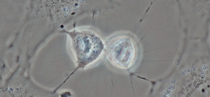
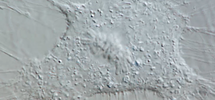
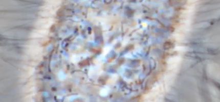
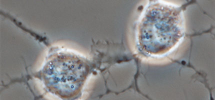
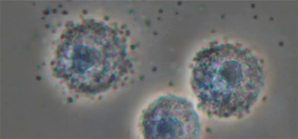
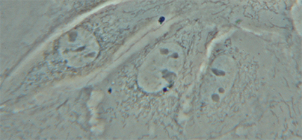
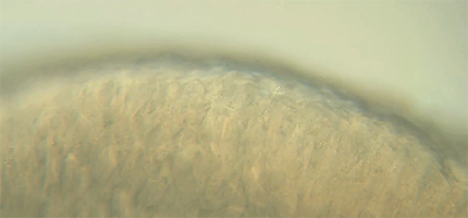
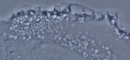
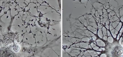
アルツハイマー病において、アミロイドβが神経細胞を傷害し細胞死を起こすと考えられています。 こちらは神経細胞にアミロイドβを添加し、コントロールの神経細胞と3日間の現象を比較した映像です。 神経細胞の末消部分から徐々に動きが止まっていくのがわかります。また、細胞の形状も不安定になっていく様子が観察されました。
In Alzheimer's disease, it is considered that amyloidβ would give nerve cells fatal damages and trigger cell death. This movie shows a gradual change of cultured neural cells after added of amyloidβ, in comparison to the control, by shooting for 3 days. It emerged that tips’ movement of the dendroid cells were gradually arrested. Also instability of the cells’ whole shape was observed.
Cells that dance at the end of cytokinesis
×
位相差顕微鏡(対物100x)で撮影した細胞分裂。中央の球体は細胞核で、その周囲に見える細くてニョロニョロしたものはミトコンドリア。分裂を起こす前、細胞は周辺の細胞との結合を外して、繊維をひきながら縮まる。核の分裂が終わると、細胞は中央で大きくくびれ、身を揺らして切り離そうとするような動きをする。両者はいったん完全に分離するが、そのあと再び一端をつなぎ合う。骨芽細胞様細胞(MC3T3-E1)を培養し、約2時間の現象をTimelapse撮影した。
Mitosis - Time lapse photography (Differential interference contrast)
×
微分干渉顕微鏡(対物100x)で撮影した細胞分裂。中央の球体が細胞核、周囲にたくさんあるニョロニョロしたものはミトコンドリア。映像の6~7秒目、核の左右にうっすらみえる放射状のものは星状体。核は凝集して染色体を形成し、中央に整列する。やがて左右から引っ張られるように染色体分裂が起こると、ほとんど同時に強いクビレが入って細胞は2つに分かれる。それからすぐに新しい核膜が現れ、細胞分裂は完了する。骨芽細胞様細胞(MC3-T3-E1)を培養し、約2時間の現象をTimelapse撮影した。
Mitosis - Time lapse photography (phase contrast)
×
位相差顕微鏡(対物100x)で撮影した細胞分裂。中央の球体が細胞核で、その周囲に見える細くてニョロニョロしたものはミトコンドリア。広がっていた細胞は、分裂期に入るとコンパクトに縮む。核膜が消失し、凝集した染色体が中央に集まって、両極に引っ張られるように二等分される。核の分裂と同時に細胞がおおきくクビれ、細胞は2つに分かれる。骨芽細胞様細胞(MC3T3-E1)を培養し、約2時間の現象をTimelapse撮影した。
マクロファージによる細菌の貪食 -Bacterial phagocytosis by macrophage-
×
マクロファージは生体内に侵入した異物や細菌を貪食して、排除する免疫細胞です。 同じように貪食する免疫細胞に好中球がいますが、マクロファージの寿命は好中球より長く、貪食能も数倍あると言われています。 また、好中球との大きな違いとして抗原提示能力を持っています。
Macrophage is one of immune cells that phagocytose and eliminate foreign bodies and bacteria invaded in a human body. There is also neutrophil in immune cells that phagocytose in the same way. But macrophage has longer life and its phagocytic ability is said to be several times more powerful than neutrophil's. Moreover, macrophage has antigen-presenting function, which makes a great difference between them.
線毛運動のHighspeed映像 -High-speed image of ciliary motility-
×
鼻腔から気道の粘膜上皮細胞表面に生えている線毛は1秒間に約15回の早さで動いて粘液とともにウイルスや細菌を体外へ排出しています。 1秒を20秒に伸ばしたHighspeedカメラで見ると、鞭をうつような線毛の動きが観察できます。この規則正しい連動した動きで線毛上部の粘液を、そこに付着した病原体とともに送り出し、感染を防いでいます。
Cancer Dance movement
×
Timelapse Vision, Inc. was awarded FIRST PLACE for our film in "Celldance 2012" The ASCB's Eighth Annual Cell Biology Video Contest.
好中球による細菌の貪食 -Bacterial phagocytosis by neutrophils-
×
好中球が細菌を貪食している様子です。 好中球は白血球のうち最も多く約7割を占める細胞です。 このように好中球は体内に侵入してきた細菌を貪食し排除することで、生体防御における重要な役割を担っています。 この仕組みは自然免疫系と呼ばれています。 貪食された細菌は食胞に取り込まれ、活性酸素によって殺菌されます。緑に光っているのが活性酸素です。
A neutrophil is phagocytizing bacteria. Neutrophils are the majority of white blood cell and they have occupied up to 70%. They play important roles in biological defense by phagocytizing and eliminating bacteria that invaded a living body, and this biological mechanism is called innate immune system. Phagocytosed bacteria are taken into the phagosome and sterilized by reactive oxygen species (ROS), emitting green fluorescence.
神経細胞 -Neuron cell-
神経細胞 -Neuron cell-
×
神経細胞は神経突起を介して他の神経細胞とつながり情報を伝達します。この培養した神経細胞もいくつもの神経突起を伸ばして隣接する神経細胞とつながり、神経ネットワークを作っています。 神経突起からはさらに細かい無数の突起が伸びているのがわかります。
マスト細胞の脱顆粒 -Degranulation of mast cells-
×
マスト細胞が、細胞質に貯蔵された顆粒を放出しています。 この現象を脱顆粒と呼びます。 マスト細胞は全身の粘膜および結合組織に存在しており、細胞膜には体外から入ってくる異物を認識するIgE抗体が多数付着しています。 それらIgEに抗原が結合することで脱顆粒が引き起こされます。 これらの顆粒にはヒスタミンなどのケミカルメディエーターが含まれ、花粉症や蕁麻疹などの即時型アレルギー反応を引き起こします。
Mast cells are releasing granules that have been stored in their cytoplasms, this phenomenon called the degranulation. They exist in mucosal and connective tissues throughout the whole body. Around the cell membrane of the mast cells, lots of IgE antibodies have adhered and they recognize foreign substances entering from the outside of the body. It is by antigens attaching to these IgE antibodies that the degranulation is triggered. These granules contain chemical mediators such as histamine and they consequently cause immediate allergic reactions—for example, hay fever and urticaria.
培養細胞 -Cultured cells-
×
シャーレで培養した細胞をタイムラプス撮影した映像です。 細胞の中に黒い核小体のある丸い核が見えます。 その周りの紐のようなものはミトコンドリアです。 顕微鏡を覗いただけでは静止しているように見える細胞も、時間をおいて1フレームずつ撮影することで細胞のダイナミックな動きを見ることができます。
鼻粘膜の線毛運動 -Nasal mucosa-
×
鼻の粘膜は線毛を持った線毛細胞に被われています。粘膜の表面を波打つように見えるのが線毛の動きです。 線毛細胞は気管や気管支にも存在し、呼吸器に入った異物を体外に排出する役目を果たしています。
Cancer Dance
Cancer Dance
×
Timelapse Vision, Inc. was awarded FIRST PRIZE for our film in "Celldance 2011" The ASCB's Seventh Annual Cell Biology Video Contest.
  • Micro

    Works

    Store
    • eミクロ
    • ぱらぱらミクロ
  • About
    • メッセージ
    • 映像制作の体制
    • 納品までの流れ
    • 表現のかたち
    • 会社概要
    • 主要取引先
    • 特定商取引法
  • Lab
    • 社内設備
    • 論文/学会発表リスト
  • News
  • Contact
    • アクセス
    • お問い合わせ

    • English
顕微鏡撮影・映像制作|株式会社タイムラプスビジョン Live imaging and Time-lapse microscopy: TIMELAPSE VISION INC.
Stock Imagery:
Copyright (C) 2023 TIMELAPSE VISION INC. All Rights Reserved.